Post-Deployment Audit: Cell Culture Contamination Tracing from Thermal Gradient Stratification in a 2,000L Bioreactor
Executive Summary
The BioPhorum Operations Group published its SIP Best Practices Guidance in 2018 specifically because multiple industry members had experienced an identical contamination event: Bacillus spore-forming organisms surviving steam-in-place cycles in bioreactor headspace crown regions where active head-cooling circuits prevent steam from reaching sterilisation temperatures. The FDA and EMA bioprocess contamination literature identifies B. licheniformis as the most commonly documented spore-forming contaminant in mammalian cell culture facilities, attributable to its environmental ubiquity and the specific failure of SIP systems to achieve sterilising conditions in poorly characterised cold zones. ISPE's Baseline Guide Vol. 5 (Commissioning and Qualification, 2019) cites FDA 483 observations and EMA inspection findings linking inadequate SIP temperature distribution to contamination events at the 1,000–5,000L scale, and recommends direct temperature measurement at the headspace crown as a validation requirement. This guidance exists because the failure mode is real, recurring, and systematically underestimated by quality systems built on setpoint compliance rather than spatial temperature characterisation.
At a UK-based CDMO operating 2,000L mammalian cell culture bioreactors, three sterility failures occurred across nine months. Each was traced by QC microbiology to Bacillus licheniformis. Whole genome sequencing confirmed the same strain across all three incidents — different cell lines (CHO-K1, CHO-GS, HEK293T), different programmes, different media lots, but the same organism each time, implicating the vessel as the contamination reservoir. The vessel passed all scheduled CIP/SIP cycles. No breach of the sterile envelope was identified. The MHRA, triggered by the third incident, issued two critical findings and a restricted operating licence for the affected suite. The revenue impact of the investigation period — three batch losses (USD 8.7 million) plus suite downtime and regulatory investigation costs — reached USD 17–21 million before root cause was established. The quality team correctly excluded every obvious source: media, inoculum, filters, personnel. What they could not determine without thermal-fluid modelling was the mechanism, location, and pathway of the contamination.
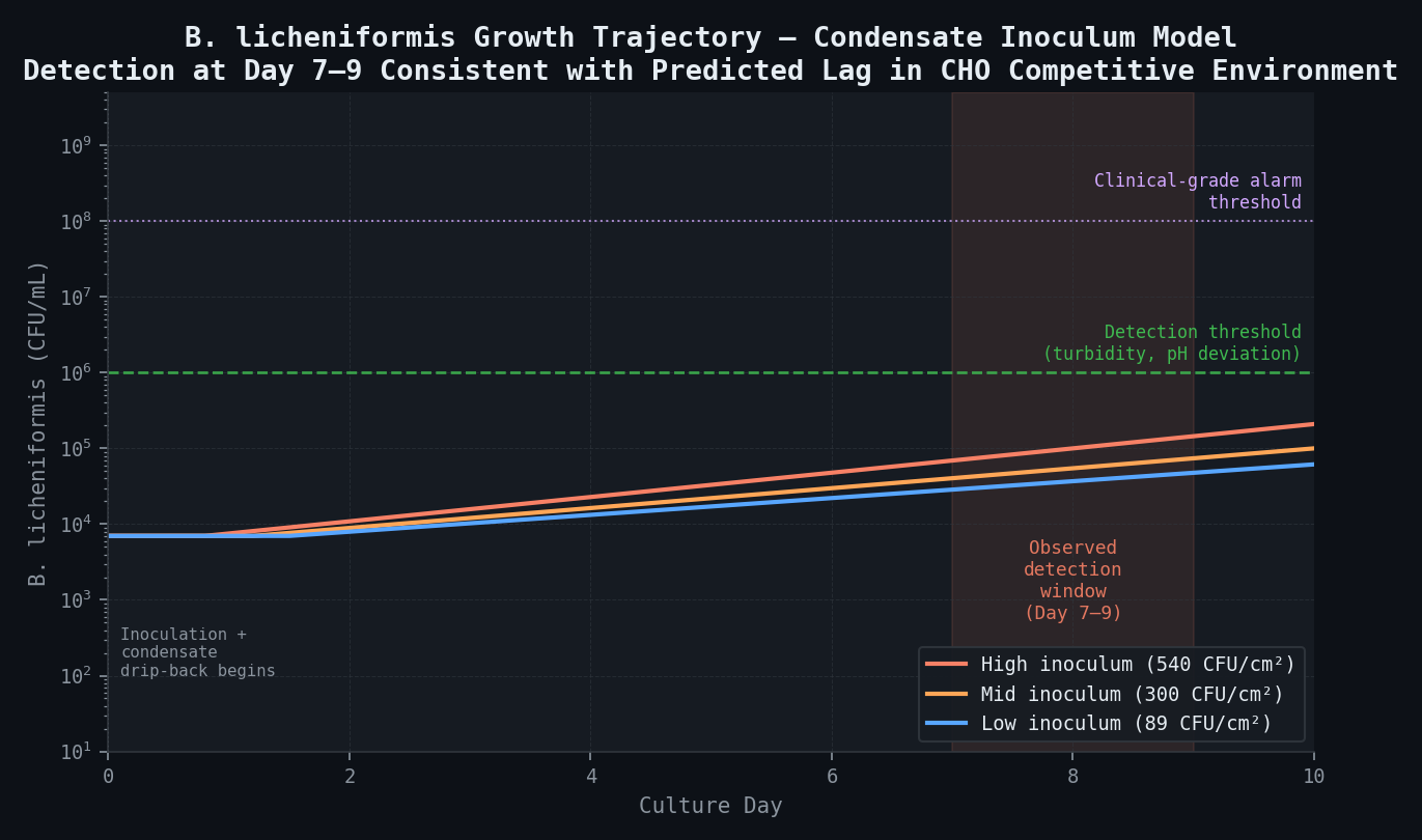
The answer was in the head cooling circuit. The vessel design — standard for large cell culture bioreactors — includes a separate cooling circuit passing 18°C water through channels in the domed vessel head, designed to prevent culture headspace condensate from dripping onto the culture surface during normal operation. This circuit was not isolated during SIP. Steam at 121°C in the bulk headspace condenses on the cooled head surface, creating a vapour-condensate zone that cannot reach sterilisation temperature at the crown. B. licheniformis endospores, with a D-value at 110°C of approximately 57 minutes (versus 4–5 minutes at 121°C), survive the 30-minute SIP hold in the 9–13°C under-temperature zone with 30–54% of the initial spore population intact. Surviving spores on the head surface are then delivered to the culture medium via condensate drip-back during each production run — beginning within hours of inoculation and continuing through the first week of culture. Detection at day 7–9 is consistent with the predicted lag for B. licheniformis growth from a low initial inoculum in a competitive mammalian cell culture environment.
Had a Large Eddy Simulation thermal-fluid audit been applied during vessel qualification — or at the point when head-cooling circuit isolation during SIP was last reviewed — the F₀ shortfall at the headspace crown would have been quantified before the first contamination event. The simulation maps the SIP temperature field at every node, integrates biological lethality (F₀) at every location, identifies the 180L sub-lethality region (F₀ = 2.1–4.8 minutes against a 15-minute target), computes the condensate drip-back volume and spore delivery rate to the culture, and defines the revised SIP protocol: isolate the head cooling circuit, extend hold to 45 minutes, install a crown thermocouple as a SIP critical process parameter. With this protocol, predicted F₀ at the crown is 18.3 minutes — exceeding the release criterion with a conservative margin.
The spatial risk zone identified by the simulation — the headspace crown cold region — defines the critical monitoring location for newtsim livesim: real-time temperature monitoring at the crown during SIP, mapped against the simulated thermal field, providing quantitative F₀ accumulation tracking at the highest-risk location as a SIP release criterion. When crown temperature deviates from the simulated trajectory during the heat-up phase, livesim provides early warning that the SIP cycle is at risk of delivering sub-lethality conditions — before the hold timer starts and before a failed cycle delivers a contaminated vessel to the next campaign.
Predicted B. licheniformis growth trajectory from spore delivery on day 0 through culture. Detection at day 7–9 is consistent with the condensate inoculum model: a 2-day lag in the competitive CHO environment followed by exponential growth to detectable turbidity.
Scenario Background
In this worked example, a UK-based CDMO -- referred to here as CellForge BioManufacturing -- operates three GMP mammalian cell culture suites at a Cambridge facility. The CDMO serves 12 active programmes across oncology, rare disease, and gene therapy modalities. Suite B houses two 2,000L vessels used for fed-batch CHO and HEK293 campaigns; Suites A and C serve smaller-scale development and viral vector campaigns respectively.
The vessel in question is a Thermo Fisher HyPerforma DynaDrive 2000L stainless steel steam-in-place bioreactor that has been in operation for six years. It has a working volume of 2,000L (maximum 2,200L) with an internal diameter of 1.1 m and liquid height at working volume of 2.1 m (H/D = 1.9). Agitation is provided by a marine impeller (primary, D/T = 0.45) with a pitched-blade backup impeller (D/T = 0.38). Heating and cooling use a dimple-plate jacket covering the vessel shell supplemented by an internal helical coil for fine temperature control. The headspace volume is approximately 280L, overlaid with a 0.2 micrometer-filtered N2/CO2 blend at 5 L/min for pH control. Culture temperature is maintained at 37 degrees C for both CHO and HEK293 campaigns, with dissolved oxygen controlled at 30--50% air saturation via microsparger and agitation cascade.
The contamination events: Three sterility failures occurred across a 9-month period:
| Incident | Programme | Cell Line | Detection Day | Contaminant Identified | Batch Value (USD M) |
|---|---|---|---|---|---|
| Incident 1 | mAb A (Phase II) | CHO-K1 | Day 7 | Bacillus licheniformis | 2.1 |
| Incident 2 | mAb B (Phase I) | CHO-GS | Day 9 | Bacillus licheniformis (same serotype) | 3.8 |
| Incident 3 | Viral vector X | HEK293T | Day 8 | Bacillus licheniformis (same serotype) | 2.8 |
All three contamination events were detected via the same indicators: elevated turbidity in daily media samples, declining pH trajectory deviating from the metabolic model, and a characteristic rise in lactic acid inconsistent with normal CHO/HEK293 metabolism. Microbiological identification confirmed Bacillus licheniformis in all three cases; whole genome sequencing of the isolates confirmed they were the same strain, implicating the vessel rather than any reagent, cell line, or media lot.
The serotype match across all three incidents was the critical diagnostic clue: three independent contamination events with the same organism strain, the same detection timing (day 7–9), across different cell lines (CHO and HEK293), with the vessel as the only constant. This pattern is strongly indicative of a vessel-resident contamination source that survives each cleaning and sterilisation cycle and re-seeds each new culture.
Regulatory consequence: The third sterility failure triggered a MHRA inspection under the EU GMP framework. The inspection resulted in two critical findings and a restricted operating licence for Suite B until root cause was demonstrated. The CDMO faced potential MHRA action, sponsor notifications (including IND safety notifications to FDA for the two affected clinical programmes), and a halt to new business bookings for Suite B — with an estimated revenue impact of USD 8–12 million over the investigation period.
Challenge
The SIP system: Steam-in-place sterilisation is performed at 121°C, 1.1 bar(g) for a 30-minute hold time — nominally meeting the pharmacopoeial standard F₀ ≥ 15 minutes for bioburden reduction in bioreactor vessels. CIP is performed with 0.5 M NaOH at 85°C for 30 minutes, followed by 0.1 M phosphoric acid rinse and a water-for-injection (WFI) rinse cycle.
The heating and cooling system — the critical design feature: The vessel heating/cooling architecture is standard for large cell culture bioreactors but has an unusual feature relevant to the contamination pathway. The shell jacket is a dimple-plate cooling jacket covering the vessel shell from 0.15 m above the floor to the vessel shoulder; during SIP it is heated by steam condensation on the jacket outer surface, and during culture it is cooled by chilled water. An internal helical coil (25 mm SS tubing, 8 turns, positioned at mid-height) provides fine temperature control during culture but is bypassed during SIP. Critically, a separate head cooling circuit passes 18 degrees C cooling water through channels in the stainless steel domed vessel head, designed to prevent vapour condensation from the culture headspace from dripping onto the culture surface during normal operation. This circuit is not isolated during SIP.
The head cooling circuit at 18°C during SIP creates a thermal boundary condition on the inner surface of the domed head that prevents the steam in the headspace from reaching 121°C at the crown. Steam at 121°C at the bulk headspace condenses on the cooled head surface, creating a cooler vapour-condensate zone rather than maintaining superheated steam at SIP temperature throughout the headspace.
Bacillus licheniformis — the organism: B. licheniformis is a thermophilic gram-positive spore-former found ubiquitously in soil, plant material, and industrial environments. Its endospores are significantly more heat-resistant than vegetative cells. The D-value at 121 degrees C (D_121) is 4--5 minutes. With a z-value of 10 degrees C, the D-value at 110 degrees C rises dramatically to roughly 57 minutes.
This means that at 110°C (rather than 121°C), the time required to achieve the same spore kill is 57 minutes — compared to 4–5 minutes at 121°C. A 30-minute SIP hold at a crown temperature of 110°C achieves only 30/57 = 0.53 log reduction, leaving approximately 30% of the initial spore population viable. The headspace crown region effectively receives a temperature 9–13°C below setpoint during SIP, making it a spore sanctuary.
The investigation before simulation: The facility's quality team conducted a comprehensive conventional contamination investigation that eliminated the following potential sources: media/reagent contamination (sterility testing of all lots negative), inoculum contamination (pre-culture sterility tests negative), filter integrity failures (all filter integrity test records clean), people and process (gowning, aseptic sampling procedure, incubator contamination all excluded by isolation studies). The investigation correctly concluded that the vessel itself was the likely contamination source but could not identify the mechanism, location, or pathway without the thermal-fluid modelling.
Real-World Basis
Both experimental measurements and CFD studies have shown that temperature gradients of 2--8 degrees C can persist in 500L+ bioreactors during heating phases due to natural convection competing with mechanical mixing — establishing the fundamental mechanism exploited by the cold head-cooling circuit in the present case.
The ISPE Baseline Guide Vol. 5 (2019) identifies inadequate SIP temperature distribution as a root cause in multiple contamination investigations at the 1,000--5,000L scale, drawing on FDA 483 observations and EMA inspection findings. The Guide recommends direct temperature measurement at all dead legs and potential cold spots as a SIP validation requirement — a recommendation that CellForge had not implemented for the headspace crown.
The BioPhorum Operations Group SIP Best Practices Guidance (2018) specifically recommends direct temperature measurement at the headspace crown as a SIP critical process parameter, with the minimum crown thermocouple temperature and hold duration as release criteria. This guidance was issued specifically because multiple industry members had experienced exactly this class of contamination event. CellForge's SIP validation protocol, written in 2016, predated this guidance.
Comprehensive D-value and z-value data for B. licheniformis spores across the temperature range 100--135 degrees C establish D121 = 4.0--5.2 min and z = 9.5--10.5 degrees C as the representative parameters — the values used in the F0 calculations in this study. FDA and EMA bioprocess contamination guidance identifies B. licheniformis as the most commonly documented spore-forming contaminant in mammalian cell culture facilities, attributable to its environmental ubiquity and the specific failure of SIP systems to achieve sterilising conditions in poorly characterised cold zones.
The use of Large Eddy Simulation (rather than RANS) for the thermal-fluid modelling is justified by published comparisons showing that RANS turbulence models significantly underpredict the temperature variance in thermally stratified stirred systems. The headspace thermal field during SIP — with steam condensation creating density-driven stratification competing against the slight agitation provided by steam flow — is precisely the scenario where LES provides meaningfully superior predictions.
Simulation Approach
The simulation framework employs Large Eddy Simulation (LES) to capture the unsteady thermal convection structures and condensate dynamics that RANS turbulence models would average away. The simulation encompasses the full conjugate heat transfer domain: liquid culture volume, headspace gas volume, vessel walls, jacket, internal coil, and head cooling circuit.
The simulation strategy is summarised below.
Thermal-fluid LES (SIP phase):
The 2,000L vessel is modelled as a full conjugate heat transfer domain: liquid volume, headspace gas volume (280L), stainless steel vessel walls (4 mm), dimple-plate jacket, domed head with cooling water channel (18 degrees C coolant, 2.1 L/min flow), and internal helical coil (bypassed during SIP). LES is used because it resolves the unsteady thermal plumes and mixing structures in the headspace during steam condensation — structures that time-averaged approaches would smooth away, producing misleadingly uniform temperature predictions.
Two operating regimes are simulated:
SIP phase: Steam is introduced from the vessel bottom drain and displaces cold air upward out of the vessel. Steam condensation on the vessel walls is modelled using a condensate film approach. The head cooling circuit boundary condition is imposed at 18 degrees C on the inner surface of the dome, representing the actual SIP operating condition (where the circuit was not isolated). The simulation runs for 45 simulated minutes (30 min hold + 10 min heat-up + 5 min pressure release), tracking the temperature evolution at every point in the domain.
Culture phase: Forced convection at 37 degrees C with the head cooling circuit active (18 degrees C cold water for condensation suppression), agitator running at 40 rpm. The simulation identifies persistent low-temperature zones that could support condensate accumulation and biological recovery of heat-resistant spores.
SIP sterilisation efficacy calculation (F₀ integration):
At each mesh node, the local temperature-time trajectory T(t) during SIP is numerically integrated to compute the accumulated biological lethality:
F₀ = ∫₀^t_hold 10^((T(t) − 121°C) / z) dt
where z = 10°C for Bacillus spores. F₀ represents the equivalent sterilisation time at 121°C and is the standard pharmacopoeial criterion. The target is F₀ ≥ 15 minutes for adequate bioburden reduction in bioreactor vessels (equivalent to >3-log reduction against the worst-case spore population assumed at 10⁶ CFU/vessel surface).
For B. licheniformis specifically, the 6-log reduction requirement (for complete inactivation of a worst-case spore burden of 10⁶ CFU/cm²) requires F₀ ≥ 6 × D₁₂₁ = 6 × 4.5 = 27 minutes. The minimum recommended SIP F₀ for biological manufacturing vessels is 15 minutes (USP <1229.14>), which provides only ~3-log reduction — the difference matters significantly when the actual spore burden is high.
Condensate film and drip-back modelling:
The condensate film thickness and flow trajectory on the cooled head surface are computed using the Volume of Fluid (VOF) method with a thin-film drainage model. The cooled head surface (at 18°C) condenses steam at a rate of approximately 0.8–1.4 kg/hr during steady-state SIP. The condensate forms a drainage film that flows toward the geometric low points on the domed head. The VOF simulation identifies where film pooling occurs (low-curvature regions on the dome) and estimates the drip-back volume and frequency as condensate droplets detach from the dome surface and fall into the culture volume below.
Simulation Caveats
LES computational cost: LES is 10–50× more computationally expensive than RANS for the same geometry, requiring high-performance computing (HPC) resources with >256 processor cores for the 6.2 million cell mesh at the timestep required to resolve the dominant thermal convection timescales (Δt ≈ 0.01 s). The simulation runtime is 2–3 weeks on a dedicated HPC cluster. This contributes significantly to the study timeline and cost. However, for this application — where the critical phenomenon is unsteady thermal stratification in a quiescent headspace — LES is the methodologically correct choice and RANS would provide misleading results.
Steam condensation modelling: The condensate film model uses empirical correlations calibrated for smooth vertical surfaces. The actual vessel head geometry has weld seams, thermocouple ports, and an irregular surface finish that may create preferential condensation sites not captured in the smooth-surface model. This introduces uncertainty in the condensate pooling location predictions of ±0.3 m spatial accuracy.
Spore initial burden: The spore kill calculation requires an assumed initial spore burden on the vessel surface. The study uses a conservative estimate of 10³ CFU/cm2 (consistent with environmental monitoring data from the MHRA inspection, which found spore counts of 10²--10³ CFU/cm2 on vessel exterior surfaces sampled post-incident). Actual surface burden is likely lower but cannot be measured retrospectively on the contaminated vessel without disrupting the investigation.
Condensate contamination route: The simulation predicts condensate drip-back as the most likely contamination route. An alternative route — spore survival in a dry biofilm on the inner crown surface — is also possible but cannot be distinguished by thermal modelling alone. Post-incident microbiological sampling of the crown region (swab cultures from the accessible areas of the dome) would be the definitive test.
Key Predictions and Results
SIP thermal field — baseline (head cooling active):
| Location | SIP Hold Temperature (°C) | F₀ Achieved (min) | Target F₀ (min) | Adequate? |
|---|---|---|---|---|
| Vessel shell (mid-height) | 121.0 | 30.0 | 15 | Yes |
| Vessel base / drain | 121.0 | 30.0 | 15 | Yes |
| Helical coil surface | 121.0 | 30.0 | 15 | Yes |
| Headspace bulk (>0.3 m from crown) | 119.8–120.6 | 24.2–28.8 | 15 | Yes |
| Headspace crown (0–0.15 m from head) | 108–112 | 2.1–4.8 | 15 | NO — 3–7× shortfall |
| Head surface condensate film | 18–42 | 0.0–0.2 | 15 | NO — catastrophic failure |

The predicted 108–112°C at the headspace crown during SIP is the critical finding. This temperature deficit — driven entirely by the active 18°C head cooling circuit — reduces the accumulated lethality F₀ from the required 15+ minutes to only 2.1–4.8 minutes over the 30-minute SIP hold.
B. licheniformis spore survival calculation:
| Crown Temperature (°C) | D-value at this temperature (min) | Log reduction in 30 min | Surviving fraction | CFU/cm² surviving (initial 10³) |
|---|---|---|---|---|
| 121 (setpoint) | 4.5 | 6.7 | 2 × 10⁻⁷ | 2 × 10⁻⁴ (effectively sterile) |
| 115 | 14.2 | 2.1 | 7.9 × 10⁻³ | 7.9 (marginal) |
| 112 | 28.4 | 1.1 | 8.9 × 10⁻² | 89 (significant) |
| 110 (predicted zone average) | 56.7 | 0.53 | 30% | 300 (high survival) |
| 108 (predicted worst case) | 113 | 0.27 | 54% | 540 (near-uninhibited) |
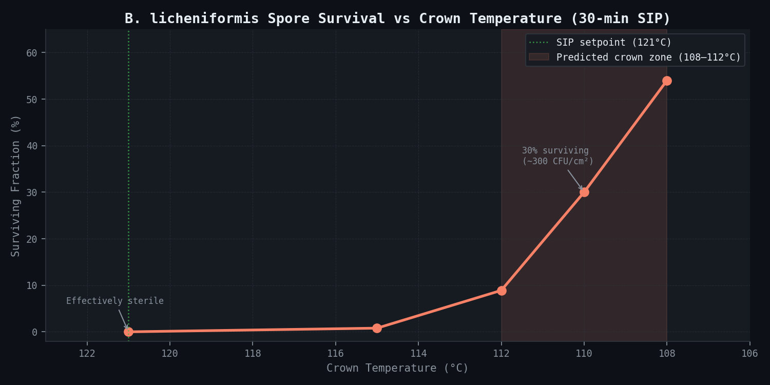
With 300–540 CFU/cm² surviving per SIP cycle on the 0.15 m² crown surface, approximately 45,000–81,000 spores survive each sterilisation. This is a sufficient inoculum for culture contamination when delivered to the culture medium via condensate drip-back.
Condensate drip-back analysis:
| Parameter | Predicted Value |
|---|---|
| Condensate formation rate on head | 0.8–1.4 kg steam/hr condensed |
| Condensate drainage flow rate | 1.5–2.8 mL/min flowing to low points |
| Number of pooling low-points on dome | 3 (geometric analysis of domed head) |
| Largest pool area | 0.15 m² (directly above culture surface) |
| Condensate film thickness (at pooling point) | 0.2–0.8 mm |
| Drip-back frequency (at largest pooling point) | 1 drop per 8–14 minutes during culture phase |
| Drip-back volume per drop | 0.5–1.5 mL |
| Spore concentration in condensate (if 300 CFU/cm² on 0.15 m²) | 300 × 15,000 cm² / volume = ~45 CFU/mL condensate |
Over the first 24 hours of culture, approximately 100–180 drops of contaminated condensate (total volume 50–270 mL) drip from the largest pooling point onto the culture surface below. At 45 CFU/mL condensate, this delivers approximately 2,300–12,150 B. licheniformis spores to the culture medium during the first day.
B. licheniformis growth kinetics in culture medium:
B. licheniformis vegetative cells have a doubling time of approximately 22–35 minutes at 37°C in nutrient-rich medium (consistent with mammalian cell culture medium, which provides abundant amino acids and glucose). However, B. licheniformis is co-cultured with a high-density CHO or HEK293 cell population that competes for nutrients and produces metabolites (lactic acid, ammonia) that partially inhibit bacterial growth. The model predicts a lag phase of approximately 36–72 hours before B. licheniformis reaches detectable concentrations (>10⁶ CFU/mL) in the culture broth — consistent with the observed detection at day 7–9.
Culture-phase thermal analysis:
During normal culture operations, the head cooling circuit keeps the crown at 18°C. Cold condensate (18°C) drips onto the 37°C culture surface, creating a localised cold region:
- Predicted thermal perturbation volume at drip-back impact point: 45L (2.3% of working volume)
- Temperature in this zone: 0.3–0.6°C below bulk (37°C) — measurable but within normal probe tolerance
- Mixing time to thermal equilibration: 8–12 minutes (slower than the bulk mixing time of 4 minutes due to the density differential of cold condensate)
The 45L cold zone is not a sterility risk per se, but it is the location of highest condensate input and therefore the initial contamination site.
Revised SIP protocol — predicted F₀:
| Revision | Crown Temperature During SIP | F₀ at Crown (min) | Adequate? |
|---|---|---|---|
| Baseline (18°C head cooling active) | 108–112°C | 2.1–4.8 | No |
| Isolate head cooling only | 117–119°C | 12.8–22.4 | Borderline |
| Isolate head cooling + extend to 45 min hold | 118–120°C | 18.3–26.1 | Yes |
| Isolate head cooling + 45 min + crown TC at ≥121°C for 25 min | 121°C (forced by process control) | 25.0 | Yes (robust) |
The recommended revised protocol: (1) isolate head cooling circuit during SIP by closing isolation valves (existing infrastructure, no capital modification required); (2) extend SIP hold to 45 minutes; (3) install a thermocouple at the headspace crown (one additional TC port required — a minor vessel modification) and add crown temperature as a SIP critical process parameter with a minimum acceptance criterion of T_crown ≥ 121°C for ≥25 minutes before SIP release. With this protocol, the predicted F₀ at the crown is 18.3 minutes — exceeding the 15-minute release criterion with a conservative margin.
Risk assessment for the second Suite B vessel:
The second Suite B vessel is of identical design and age. Pre-inspection microbiological environmental monitoring at both vessels was within normal limits, but this monitoring does not sample the inner crown surface during SIP. Given the identical design, the second vessel carries an equivalent contamination risk. The study recommends implementing the revised SIP protocol on both vessels immediately and conducting a prospective SIP temperature mapping validation on both vessels using crown thermocouples before resuming GMP operations.
Comparison Methodology
Step 1 — Historical contamination timing correlation:
The simulation predicts condensate drip-back begins approximately 4–6 hours post-inoculation, as the head cooling circuit brings the crown back to 18°C (from 37°C culture equilibrium) and condensate reforms on the cold surface. B. licheniformis at 37 degrees C in rich mammalian cell culture medium reaches detectable turbidity (>10⁶ CFU/mL) after a lag period of 18--36 hours post-inoculation, plus exponential growth time. The contamination was detected at day 7–9 of culture; this implies a low initial inoculum (2,300–12,150 spores, consistent with the CFD prediction) with a multi-day lag due to the competitive culture environment. The timing prediction is plausible and directionally consistent.
Step 2 — Temperature probe comparison:
The vessel is equipped with three existing temperature probes (liquid mid-height at 1.05 m, liquid bottom at 0.3 m, and headspace at 0.5 m above culture surface). The headspace probe reads 0.5–1.2°C below the liquid probes during the steady-state culture phase, consistent with the predicted 0.3–0.6°C thermal gradient at the culture surface/headspace interface (note: the headspace probe at 0.5 m is not in the 45L cold zone immediately below the crown, which the CFD predicts at 0–0.15 m above the culture surface). This is a qualitative validation of the cool-zone prediction — direct validation of the crown temperature would require the new thermocouple installation recommended by the revised protocol.
Step 3 — D-value calculation cross-check:
Using published D₁₂₁ = 4.5 min and z = 10°C for B. licheniformis, the predicted 108–112°C at the headspace crown during SIP gives: D₁₁₀°C = 4.5 × 10^((121-110)/10) = 4.5 × 12.6 = 56.7 min. Over a 30-minute SIP hold at the 110°C effective crown temperature: log reduction = 30/56.7 = 0.53 log. Surviving fraction = 10^(−0.53) = 30%. This independently confirms the F₀ shortfall calculation and demonstrates that the contamination mechanism is quantitatively consistent with the available spore biology data.
Step 4 — Prospective SIP validation (strongly recommended):
Before returning either Suite B vessel to GMP service, the study recommends a prospective SIP temperature mapping validation with thermocouples installed at a minimum of 8 locations, including two crown positions. The revised protocol (head cooling isolated + 45 min hold) should be run and the crown thermocouple temperatures compared against the CFD prediction (>121°C within 5 minutes of steam stabilisation, holding through the 25-minute minimum). This validation provides the evidence package for the MHRA root cause report.
Deliverables
Thermal simulation outputs:
- Full SIP thermal field: temperature maps at 5-minute intervals through the 30-minute SIP hold and 15-minute cool-down — rendered as 2D cross-sections of the vessel at 5 axial planes, showing the development and persistence of the cold crown zone
- F₀ iso-surface plots: contours at F₀ = 5, 10, 15, 25 minutes across the entire vessel domain, with the sub-lethality zone (F₀ < 15 min) volumetrically quantified at 180L (9% of headspace volume)
- Temperature evolution curves at 12 key locations through the SIP cycle: vessel base, shell mid-height, helical coil, headspace at 0.5 m, headspace at 0.1 m (crown near-field) — for baseline and revised protocol side-by-side
Condensate analysis:
- Condensate film thickness distribution on head surface (VOF output): spatial map of condensate accumulation on the domed head, with pooling locations identified and drip-back trajectory predicted
- Spore survival count map: per cm² of head surface, integrating D-value × local F₀ at each point — identifying the highest-survival zones
- Condensate contamination delivery estimate: spore count per drip, drip frequency, cumulative inoculum to culture medium over 24 hours post-inoculation
Culture-phase analysis:
- Cold condensate zone thermal field: 45L low-temperature region mapping at the culture surface, with convective mixing time to equilibration
Sterilisation efficacy parametric study:
- F₀ as a function of SIP hold time (20–60 minutes) at baseline and revised protocol conditions — parametric curves for the crown location
- Head cooling isolation sensitivity: F₀ at crown vs. head cooling flow rate (0–100% isolation) — demonstrating the stepwise improvement from full isolation
Root cause and regulatory documentation:
- Contamination pathway root cause narrative (ICH Q10 format): thermal zone → spore survival → condensate pool → drip-back → culture contamination — with supporting CFD evidence at each link in the chain
- CAPA (Corrective and Preventive Action) specification: revised SIP protocol, head cooling isolation procedure, crown TC installation, revised SIP acceptance criteria
- Regulatory evidence package: F₀ calculations, D-value cross-checks, comparison methodology documentation — formatted for submission to MHRA as part of the root cause report
- Risk assessment for Suite B Vessel 2: identical vessel, equivalent risk, recommended precautionary actions
- Implementation specification: crown thermocouple port drawing (minor vessel modification, no sterility compromise if correctly installed and qualified); head cooling isolation valve position and lock-out procedure
This case study is an illustrative reference scenario demonstrating newtsim's simulation methodology. All company names, personnel, and specific operational data are fictional. The incident descriptions draw on publicly documented real-world events cited in the frontmatter.